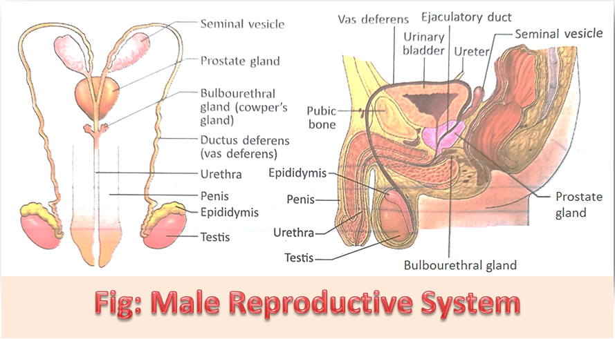
Male Reproductive System:
Different sexual organs of males which are involved in the process of production and secretion of sperms are united to form a system called the male reproductive system.
1. Scrotum:
The scrotum is a pouch of deeply pigmented skin divided into two compartments each of which contains one testis, one epididymis, and the testicular end of a spermatic cord. It lies below the symphysis pubs and behind the thigh. The coat of the scrotum consists of 7 layers which are – i. Skin, ii. Tunica dartos, iii. External spermatic fascia, iv. Cremasteric fascia, v. Internal spermatic fascia, vi. Tunica vaginalis vii. Visceral layer and viii. Parietal layer.
The scrotum maintains a temperature lower than the normal (37°C) body temperature. The normal temperature of testes in the scrotum is lower than the internal body temperature by about 2-2.5°C. This temperature is ideal for sperm development. When exposure to cold occurs the dartos muscle present in the scrotum contracts and puts up a thicker investure around the testes. It also a movement towards the pelvic cavity allows the testes to absorb the heat from the rest of the body, thus keeping the sperm warm. The inguinal canal helps the scrotum to remain connected with the pelvic cavity.
2. Testes:
The testes are the primary reproductive organs of the male and are the equivalent of the ovaries in the female. During the early fetal stage, it develops in the abdominal cavity exactly just below the kidney. In embryonic life, the testes remain within the pelvic cavity. then it gradually begins to descend through the inguinal canal and enter the scrotum about 2 months before the birth of a child. Failure of the testes to descend into the scrotum is called cryptorchidism.
3. Epididymis:
It is a convoluted tube, 6 meters in length. It remains coiled up with the intertwining connective tissue surrounding the tunic at the back of the testis. It is continued as vas deferens. The anterior end of the testes is called caput epididymis, in which vasa efferentia open. The middle part is known as corpus epididymis and the posterior part is called cauda epididymis. The secretions of epididymis provide nourishment for spermatozoa and help them to mature as sperm.
4. Spermatic Cords:
There are two spermatic cords, one leading from each testis. The spermatic cord suspends the testis in the scrotum. The spermatic cord passes through the inguinal canal. At the deep inguinal ring, the structures within the cord diverge. Each spermatic cord is composed of the testicular artery, testicular venous plexus, lymph vessels, vas deferens, and nerves. All of these are covered by a thin layer of fibrous tissue and an inner layer of muscle tissue.
5. Vas deferens:
It is the finely coiled tubule connected to the end part of the epididymis. It enters the pelvic cavity through the inguinal canal and forms a loop around the urinary bladder and opens at the base of the ejaculatory duct. Through vas deferens, sperms reach the urethra from the seminal vesicle. Vas deferens serve as a secondary storehouse for spermatozoa, which will release sperm at the time of ejaculation.
6. Seminal vesicles:
There are two seminal vesicles that lie on each side of the front part at the neck region of the urinary bladder. They are approximately 5cm long and pyramidal in shape.
7. Ejaculatory duct:
It is a fine duct through which a seminal vesicle opens into the urethra. This duct is formed by the union of the duct of the seminal vesicle and the end part of the vas deferens. Each duct is about 2 cm long. Through this ejaculatory duct, sperms are discharged with semen into the urethra during intercourse.
8. Prostate gland:
This gland lies around the urethra at the base of the neck region of the urinary bladder and is covered by a vascular capsule. It is composed of fibro-elastic tissue, mucosal gland, and plain muscles. The secretion of this gland makes the urethra slippery for the discharge of semen. It acts as a nutritive fluid for male gametes. It also increases the volume of semen. The prostatic fluid contributes 20% of the total volume of semen.
9. Cowper’s gland:
There is a pair of pea-shaped glands below the prostate gland. The glands open into the urethra through fine ducts. Cowper’s glands secrete a thin milky fluid that forms 30% volume of the semen. The secretion of these glands makes the urethra slippery.
10. Penis:
It is the muscular copulatory organ in males. It contains 3 cylindrical bodies of erectile cavernous tissue. i. Corpus spongiosum muscle at the center, ii. Corpus cavernosal muscle surrounds the corpus spongiosum from both sides.
The penis is covered by skin and is made of three parts: i. Skin, ii. Body and iii. Glans. At the anterior part of the glans, the skin is connected by fine folds. This is called a prepuce. The penis becomes hard and enlarged during intercourse.
11. Uretha:
The male urethra provides a common pathway for the flow of urine and semen. It is much longer than in females, measuring about 19-20 cm. It has three parts. The first part is surrounded by the prostate gland and is called the prostatic urethra. The second part is the membranous urethra which is situated in the perineum. The third part is the spongy urethra which is situated in the penis.