Female Reproductive System:
The different sexual organs of females which are involved in the production of ovum, process of ovulation, and fetus formation are united to form a system called female reproductive system.
1. Ovary:
There are a pair of ovaries situated in the pelvic cavity one on either side of the uterus near the free end of the fallopian tube. They are attached to the back of the broad ligament by a fold of the peritoneum called mesovarium. The ovaries are solid ovoid or bean-shaped bodies. Each ovary is 2-4 cm in length and 1.5-2.0 cm in thickness. Each ovary weighs 4-6 gm. Ovaries produce ovum and hormaones.
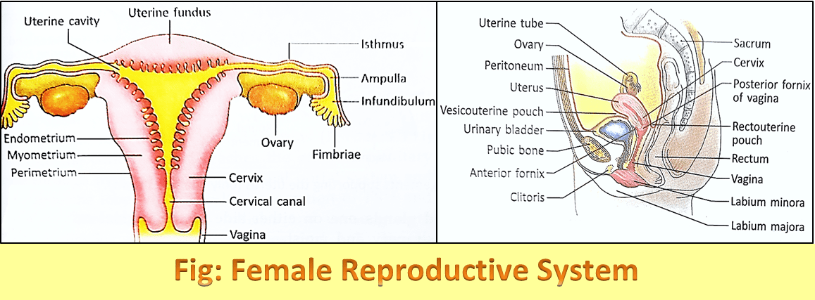
2. Fallopian tube:
These are two tubes formed from both sides of the uterus and extend up to the ovary. Each tube is 10-12 cm in length and is divided into four parts –
-
i. Uterine: It lies within the wall of the uterus between the fundus and the body.
ii. Isthmus: It is the straight narrow part just lateral to the wall of the uterus.
iii. Ampulla: It is the widest part of the tube and is long and tortuous.
iv. Infundibulum: It is a dilated trumpet-like portion opening in the peritoneal cavity. The end of the infundibulum is like a funnel and has finger-like projections. These are called fimbriae. The fallopian tube carries the ovum which is fertilized in the ampulla.
3. Uterus:
It is a pear-shaped muscular part at the middle of the urinary bladder and returns to the pelvic cavity. In adult females, it measures 6-7 cm in length and 5 cm in breadth. It is divided into 3 parts – i. Fundus, ii. Body, iii. Cervix
The walls of the uterus consist of three different types of tissue layers. The outer covering is called perimetrium. The middle thick layer of smooth muscle fiber is called the myometrium and the inner glandular layer is called the endometrium.
4. Vagina:
It is a tubular part after the cervix. it is about 8 cm in length. It opens to the exterior through the vagina orifice. The vaginal orifice is covered by the hymen. It is a stretchable organ. The acidic environment of the vagina protects it from the growth of various pathogenic organisms in it.
5. Bartholin’s gland:
These are two pear-shaped glands one on either side of the vaginal orifice. These glands secrete mucous which makes the vaginal orifice slippery and moist.
6. Vulva:
Vulva is the outer part of the female genitals. It includes the opening of the vagina so, sometimes it is called the vestibule.
7. Mammary glands:
These are two skin glands formed by the invagination of the surface epithelium into the underlying connective tissue on both sides of the chest. In males, they are small and rudimentary. Females are immature in childhood. At puberty, they become mature and well-developed under the influence of estrogen and progesterone hormones. During pregnancy, they are more enlarged.
Mammary glands are enlarged modified sweat glands. It is an elevated structure present over the pectoral region. This gland is covered by skin and underlying it discrete masses of glandular tissue are present along with connective tissue and adipose tissue. Glandular tissue consists of alveoli having secretory cells.
Nearly at the middle of each mammary gland, there is a nipple. Around the nipple a light black colored pigmented layer, the areola is present. The layer becomes dark during pregnancy. The mammary glands are made of fibrous tissue, adipose tissue, and modified muscle fibers called myoepithelium. Each mammary gland consists of 15-20 lobes. Each lobe is divided into many tubules which contain numerous ducts. The ducts branch out into terminal tubules which end in alveoli that secrete milk.