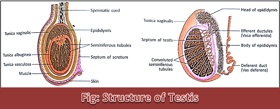
Structure of Testis:
Each adult testis is made up of the following parts:
1. Tunica albuginea:
It is the outer covering of the testis which is made of dense rigid white fibrous tissue. From tunica albuginea, a number of trabeculae enter the matrix of the testis and divide it into 100-200 pyramidal lobules.
2. Seminiferous tubules:
In each lobule of the testis, there are a number of coiled tubules covered by basement membrane and connective tissue. These are called seminiferous tubules. The combined length of each tubule is about 200-500 mm and 0.25 mm is the diameter.

3. Interstitial cells of Leydig:
These are large polyhedral cells inside the stroma in between the seminiferous tubules in the matrix of the testis. The cells are 15-20 µm in diameter and contain round fat granules. These cells secrete testosterone hormone.
4. Tubules:
There are mainly three types of tubules:
i. Straight tubule: These tubules are formed by the union of seminiferous tubules with one another. They are made of isohedral cells.
ii. Rete testis: It is formed by the union of straight tubules. They are made up of cubical epithelial cells.
iii. Vasa efferentia: These are fine tubules that arise from the rete testis and enter the epididymis. These tubules are 4 cm in length and 0.5 mm in diameter. They are made of ciliated columnar epithelial cells. The cilia help the spermatozoa to reach the seminal vesicle.
Functions of Testis:
The testis is a mixed gland and performs both (1) Exocrine and (2) Endocrine Functions.
1. Exocrine Function: It forms spermatozoa in the seminiferous tubules. This occurs under the influence of GTH of the anterior pituitary gland.
2. Endocrine Function: The interstitial cells of Leydig form and secrete the male hormone, testosterone.
Structure of Ovary:
The structure varies in childhood, puberty, pregnancy, and menopause of female life. A mature ovary is made of the following parts:
1. Germinal epithelium:
It is the outer covering layer of the ovary connected to the peritoneum mainly it is covered by the visceral peritoneum. It is made of a single layer of cubical epithelial cells. The germinal epithelium gives rise to primordial follicles.
2. Tunica albuginea:
It is the layer below the germinal epithelium. It is made of acidophilic white fibrous tissue with connective tissue cells. It performs the framework of the ovary.

3. Stroma:
It is made of a network of connective tissues attached to the tunica albuginea. It contains involuntary muscle cells tapering at both ends, blood vessels, lymphatics, and nerves.
4. Primordial follicles:
These are immature follicles formed from the germinal epithelium. Mainly, it is covered by the visceral peritoneum. They remain scattered like islands in the stroma of the ovary. In newborn babies, they are 350000 – 500000 in number which decreases with age. Each primordial follicle is made of many layers of cells without a fluid-filled cavity in between them. The primordial follicles mature into Graafian follicles.
5. Graafian follicles:
These are matured follicles in the ovary. These are formed from the primordial follicles under the influence of FSH of the anterior pituitary gland.
6. Corpus luteum:
The ruptured follicle after ovulation, is filled up by cells with coagulated blood drop and transformed into a gland. This is called the corpus luteum. It is made of large pyramidal cells and contains yellow pigment, lutein.
The corpus luteum is a temporary gland. If pregnancy occurs due to fertilization of the ovum, it persists for 3-4 months to supply estrogen and progesterone hormones to the developing embryo. Otherwise, it degenerates on every 28th day and comes out with menstruation.
7. Interstitial cells:
These are polyhedral cells in the stroma of the ovary. These cells contain numerous fat granules.
Functions of Ovary:
1. Exocrine Function: The ovary forms an ovum in the Graafian follicle.
2. Endocrine Functions: The ovary produces and secretes (i) Oestrogen, (ii) Progesterone, and (iii) Relaxin hormones.