Embryo Formation:
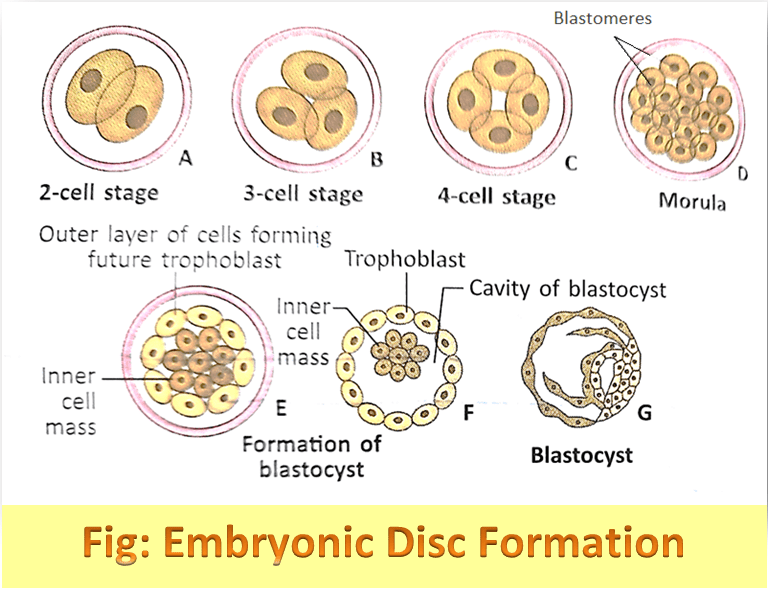
From conception til the end of the 8th week (2nd month) the organism found in the uterus is known as embryo. Through various stages, the morula transforms into an embryo (Morula → Blastula →Gastrula → Embryo).
Embryonic Disc Formation:
As we know that human blastocyst consists of trophoblast and inner cell mass. The stem cells which have the capacity to give rise to organs and all tissues are present in the inner cell mass. After 8 days of fertilization, the cells of the inner cell mass differentiate into two layers –
- Hypoblast
- Epiblast

The Hypoblast or primitive endoderm is a columnar cell layer. The epiblast or primitive ectoderm is a cuboidal cell layer. The hypoblast and epiblast cells combinedly form a two-layered embryonic disc. The three germ layers which constitute the embryonic disc are –
-
1. Endoderm
2. Ectoderm
3. Mesoderm
Endoderm and Ectoderm Formation:
Some inner cells of blastocyst lie on its free surface, which constitutes the first germ layer or endoderm. After a rapid increase in cell number a complete second layer is formed inside the original outer layer of the blastocyst. At that time embryo just look like a tube enclosing another tube of a smaller diameter. Endoderm when internally bounded by the inner tube with a lumen, it is known as the primitive gut.
At the late stage of development, the primitive gut differentiates into two portions – i. One of them is present in the body of the embryo which constitutes the gut tract, ii. Another one is known as the yolk sac, which communicates with the gut of the embryo.
After endoderm formation, the remaining cells of the inner cell mass form a cuboidal cell layer, which is called ectoderm.
Mesoderm Formation:
After endoderm establishment as a distinct cell layer mesoderm formation occurs. This mesoderm is called extra embryonic mesoderm as it lies outside the embryonic disc. As the extraembryonic mesoderm lies in between the trophoblast and flattened endodermal cell lining the yolk sac, it separates the two. It does not give rise to any tissue of the embryo itself. This extra embryonic mesoderm is differentiated into two parts the partial or somatopleuric extra embryonic mesoderm and visceral or inner splanchnopleuric extra embryonic mesoderm. Both these layers enclose the extraembryonic coelom.