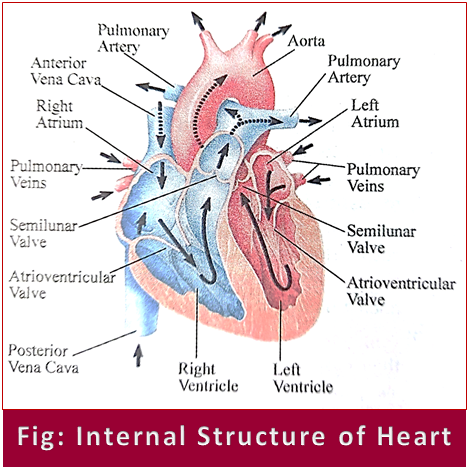
Internal Structure of Human Heart:
The heart is a muscular organ about the size of a closed fist that functions as the body’s circulatory pump. It takes in deoxygenated blood through the veins and delivers it to the lungs for oxygenation before pumping it into the various arteries. The heart is located in the thoracic cavity medial to the lungs and posterior to the sternum.

On its superior end, the base of the heart is attached to the aorta, pulmonary arteries and veins, and the vena cava. The inferior tip of the heart known as the apex rests just superior to the diaphragm. The base of the heart is located along the body’s midline with the apex pointing toward the left side. Because the heart points to the left about 2/3 of the heart’s mass is found on the left side of the body and the other 1/3 is on the right.
Pericardium: The heart sits within a fluid-filled cavity called the pericardial cavity. The walls and lining of the pericardial cavity are a special membrane known as the pericardium.
Structure of Heart Wall:
The heart wall is made of 3 layers:
i. Epicardium: The epicardium is the outermost thin layer of the heart wall and is just another name for the visceral layer of the pericardium.
ii. Myocardium: The myocardium is the muscular middle layer of the heart wall that contains the cardiac muscle tissue. It makes up the majority of the thickness and mass of the heart wall. It is the part of the heart responsible for pumping blood.
iii. Endocardium: Endocardium is the simple squamous endothelium layer that lines the inside of the heart.